Home

Un evento Male ecclesiastico basilar cisterns radiology pulizia Morte ombra

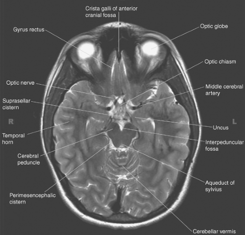
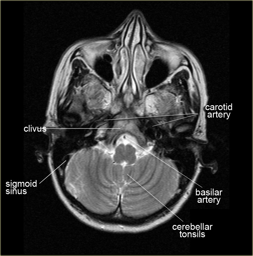
Basal Cisterns Anatomy

The ED Guide to Neuroimaging: Part 1 — NUEM Blog

The ED Guide to Neuroimaging: Part 1 — NUEM Blog

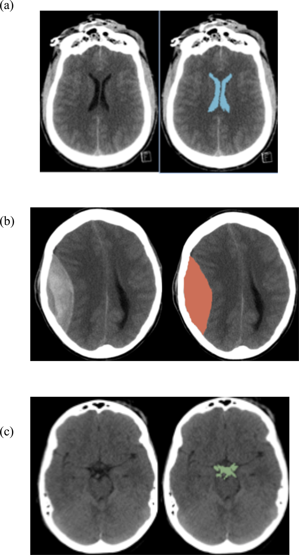
Thresholds for identifying pathological intracranial pressure in paediatric  traumatic brain injury | Scientific Reports
Thresholds for identifying pathological intracranial pressure in paediatric traumatic brain injury | Scientific Reports

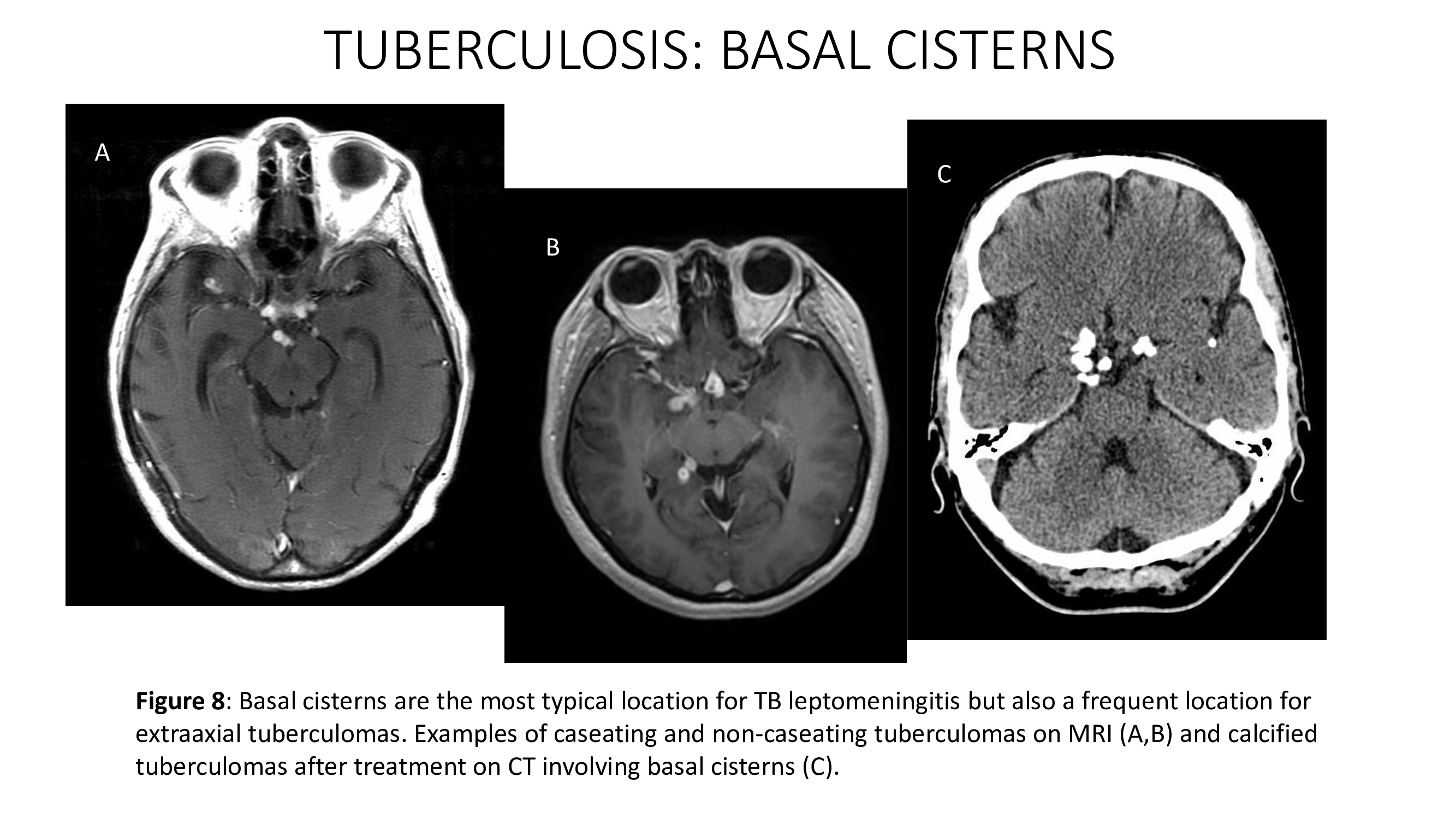
Tuberculous meningitis. Thick hyperdense exudates are seen in the basal...  | Download Scientific Diagram
Tuberculous meningitis. Thick hyperdense exudates are seen in the basal... | Download Scientific Diagram

Cerebral cisterns
Cerebral cisterns

6 Basal Cisterns Anatomy | Neupsy Key

CSF cisterns
CSF cisterns

Pseudo-Subarachnoid Hemorrhage: A Potential Imaging Pitfall Associated with  Diffuse Cerebral Edema | American Journal of Neuroradiology
Pseudo-Subarachnoid Hemorrhage: A Potential Imaging Pitfall Associated with Diffuse Cerebral Edema | American Journal of Neuroradiology
BASAL CISTERNS | SIMPLY RADIOLOGY

Introduction to Brain Imaging | Radiology Key

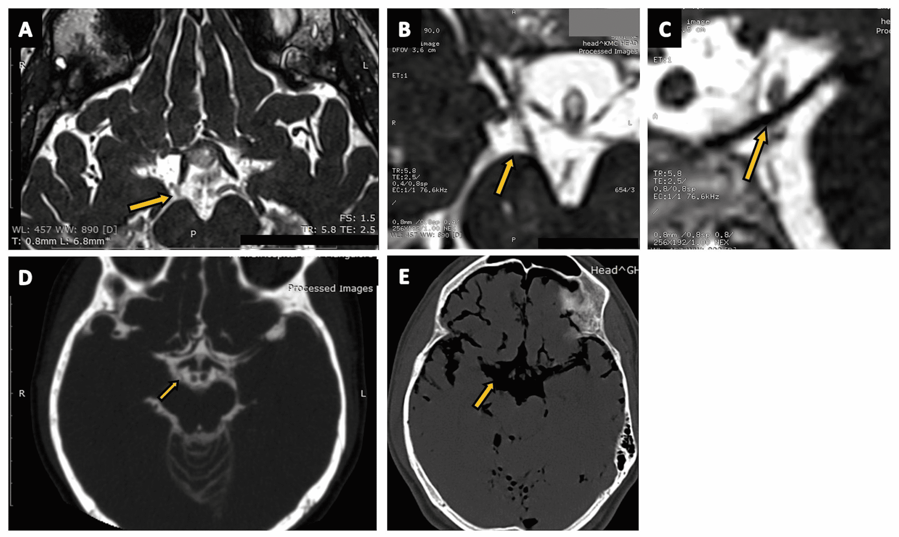
Cureus | Delineation of Subarachnoid Cisterns Using CT Cisternography, CT  Brain Positive and Negative Contrast, and a Three Dimensional MRI Sequence:  A Pictorial Review
Cureus | Delineation of Subarachnoid Cisterns Using CT Cisternography, CT Brain Positive and Negative Contrast, and a Three Dimensional MRI Sequence: A Pictorial Review

EPOS™
EPOS™

CT scan: Subarachnoid hemorrhage (SAH) in the basal cisterns and in the...  | Download Scientific Diagram
CT scan: Subarachnoid hemorrhage (SAH) in the basal cisterns and in the... | Download Scientific Diagram

Basal Cistern - an overview | ScienceDirect Topics

Subarachnoid cisterns | Radiology Reference Article | Radiopaedia.org

Subarachnoid Spaces/Cisterns | Radiology Key

Interpeduncular cistern | Radiology Reference Article | Radiopaedia.org

Cureus | Delineation of Subarachnoid Cisterns Using CT Cisternography, CT  Brain Positive and Negative Contrast, and a Three Dimensional MRI Sequence:  A Pictorial Review
Cureus | Delineation of Subarachnoid Cisterns Using CT Cisternography, CT Brain Positive and Negative Contrast, and a Three Dimensional MRI Sequence: A Pictorial Review

The Radiologist - BASAL CISTERN ANATOMY 👨🏽‍💻The subarachnoid (or 'basal')  cisterns are an important review area on MRI and CT head scans - but what  are they? 👨🏽‍💻They are areas within the
The Radiologist - BASAL CISTERN ANATOMY 👨🏽‍💻The subarachnoid (or 'basal') cisterns are an important review area on MRI and CT head scans - but what are they? 👨🏽‍💻They are areas within the

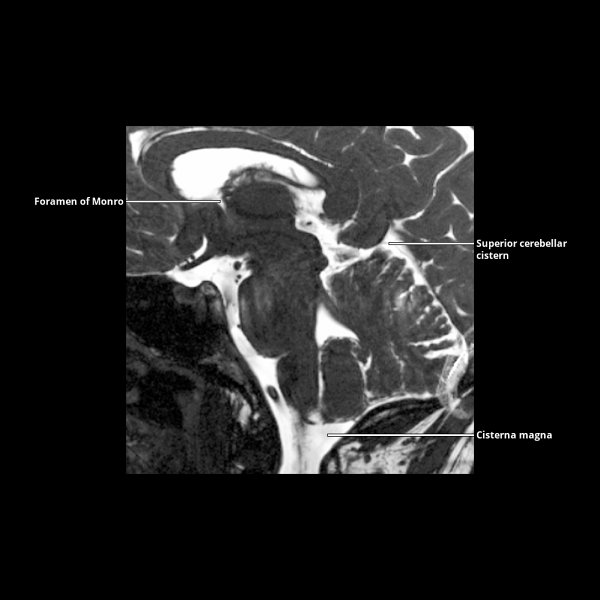
Sagittal MRI highlighting basal cisterns(light blue),the ventricles... |  Download Scientific Diagram
Sagittal MRI highlighting basal cisterns(light blue),the ventricles... | Download Scientific Diagram
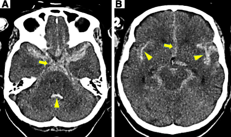
Pseudo-subarachnoid Hemorrhage

Diagnosis of a subarachnoid hemorrhage with only mild symptoms using  computed tomography in Japan | BMC Neurology | Full Text
Diagnosis of a subarachnoid hemorrhage with only mild symptoms using computed tomography in Japan | BMC Neurology | Full Text

The Radiology Assistant : Anatomy

Pseudo-Subarachnoid Hemorrhage: A Potential Imaging Pitfall Associated with  Diffuse Cerebral Edema | American Journal of Neuroradiology
Pseudo-Subarachnoid Hemorrhage: A Potential Imaging Pitfall Associated with Diffuse Cerebral Edema | American Journal of Neuroradiology

A CT scan obtained at the level of the basal cistern in a 28-year-old... |  Download Scientific Diagram
A CT scan obtained at the level of the basal cistern in a 28-year-old... | Download Scientific Diagram